
Research Article
Identification of Cutaneous Leishmaniasis in Humans Through Polymerase Chain Reaction (PCR)
Ismail Qureshi1, Mustafa Solangi2, Ahad Soomro3, Waseem Baloch1, Sundesh Kumar1, Shahjahan Qaim Khani1
1Faculty of Animal Husbandry and Veterinary Sciences, Sindh Agriculture University, Tandojam, Sindh-Pakistan; 2Vaccine Production Unit (VPU) Tandojam, Sindh-Pakistan, 3Central Veterinary Diagnostic Laboratory Research and Diagnosis (CVDL) Tandojam, Sindh-Pakistan.
Abstract | To establish the baseline and the data on samples of Cutaneous Leishmaniasis were collected from three different districts of Sindh-Province, Pakistan to diagnose Leishmania species (spp), parasites through Polymerase Chain Reaction (PCR). Total, 70 human Leishmaniasis tissues from various villages of district, Dadu, Jamshoro and Hyderabad were symptomatically identified. For further confirmation all identified tissue samples were subjected to Polymerase Chain Reaction (PCR) using, Applied Bio-system thermal cycler. DNA from samples was extracted, quantified, through, Nano drop spectrophotometer and, Polymerase Chain Reaction (PCR) product was run on, Gel electrophoresis and bands were analyzed using, Gel documentation system out of, 70 suspected, Cutaneous Leishmaniasis cases, 61 were diagnosed clinically cases and confirmed by, Polymerase Chain Reaction (PCR) for, Cutaneous Leishmaniasis. During this study, both dry and wet types of lesions were observed that, indicated the presence of two strain of parasite. Children and adults between 08-60 years of ages were found more susceptible to infection when compared with above 60 years of ages. The strains were found to be Leishmania tropica and Leishmania major as was indicated by the nature of lesions and confirmed by Polymerase Chain Reaction (PCR). Cutaneous Leishmaniasis is endemic and increasing in the rural areas of Sindh province, Pakistan. This requires attention of health authorities to take appropriate measures for its effective control, failing which it may create more severe public health problems.
Keywords | Cutaneous Leishmaniasis, Sand fly, Leishmania tropica, Leishmania major, PCR, Pakistan.
Editor | Muhammad Imran Rashid, Department of Parasitology, University of Veterinary and Animal Sciences, Lahore, Pakistan.
Received | December 24, 2018; Accepted | February 06, 2019; Published | February 28, 2019
*Correspondence | Muhammad Ismail Qureshi, Faculty of Animal Husbandry and Veterinary Sciences, Sindh Agriculture University, Tandojam, Sindh-Pakistan; Email: [email protected]
Citation | Qureshi I, Solangi M, Soomro A, Baloch W, Kumar S, Khani SQ (2019). Identification of cutaneous leishmaniasis in humans through polymerase chain reaction (pcr). J. Adv. Parasitol. 6(1): 7-11.
DOI | http://dx.doi.org/10.17582/journal.jap/2019/6.1.7.11
Copyright © 2019 Qureshi et al. This is an open access article distributed under the Creative Commons Attribution License, which permits unrestricted use, distribution, and reproduction in any medium, provided the original work is properly cited.
INTRODUCTION
Cutaneous Leishmaniasis (CL) is caused by haemo-flagellate genus Leishmania, which infect domestic and wild mammals, including human being (Gradoni et al.,1999) Cutaneous Leishmaniasis in Pakistan has been a burning seasonal problem in the Canine and Human being. Present study was therefore conducted to generate baseline data on Leishmaniasis in three districts of Sindh-Province, Pakistan (Al-Samarai et al., 2009; Aneela et al., 2011; Bari et al., 2011). Leishmaniasis is an endemic disease in certain areas of Pakistan and infection transmitted via the bite of phlebotomine sandfly and presence of its vector is associated with occurrence of cases in endemic areas, (Al-Zahrani et al., 2004). Cutaneous Leishmaniasis has emerged as a challenging infectious disease in the form of new outbreaks in different areas of Sindh-Province, Pakistan and Balouchistan, (Cortes et al., 2004; Kakar, and Suleman. 2004). This disease is wide spread and may cause serious health problems in communities through Mediterranean regions and the Middle East, including Pakistan, (Brooker et al., 2004). There are estimated 12 million cases world wide and 1.5 million new cases of Cutaneous Leishmaniasis (CL) are added each year suspected from tissue samples, (Parviz et al., 2008; Moradi., 2009). The disease affects all ages groups of human beings including children. This study was conducted to detect Leishmaniasis through clinical way and confirmed by Polymerase Chain Reaction (PCR) in suspected tissue samples.
MATERIALS AND METHODS
Study Area
Tissue samples were collected from Hyderabad were (18), Dadu (41), and Jamshoro (10) total 70 samples were collected of patients from different health centers and clinics of challenged districts in Sindh-Province, Pakistan for detection of Cutaneous Leishmaniasis (CL) via clinical diagnosis followed by Polymerase Chain Reaction (PCR).
Sample Size
Tissue samples of human beings between 08-60 years of ages were collected from ulcerative areas washed thoroughly with 70 percent alcohol and size were 0.3-0.5 cm. The tissue samples were brought at Molecular & Parasitology Laboratory, Sindh Agriculture University, Tandojam and kept them into bijou bottles that containing 3-5 ml of 70 percent alcohol to keep tissue for further processing.
DNA Extraction
DNA was extracted through Qiagen column kit. Protocol was followed according to manufactures guide.
DNA Purification Protocol
Name of kit: DNeasy Blood & Tissue Kits.
DNA Quantification
DNA was quantified on Nano drop Spectrophotometer, (ND-1000, Thermo, and Scientific, USA) at 260/280 nm wave length.
PCR Conditions
The following thermal conditions as initial denaturation at 940C for 5 minutes followed by 30 cycles including denaturation at 940C for 35 seconds annealing at 600C for 35 seconds, extension at 720C for 45 seconds and final extension at 720C for 5 minutes were used. At the end 5µl of the reaction mixture was analyzed by 2% Agarose gel electrophoresis. These primers were evaluated with Leishmania standard species including Leishmania, (MCAN/IR/97/LON 490), Leishmania major, (MHOM/IR/75/ER) and Leishmania tropica, (MHOM/IR/04/Mash 10).
Primers
Specific oligonucleotide primer as forward (5’-CAACACGCCGCCTCCTC TC T-3) and Reverse primer (5’-AAA CAA AGG TTG TCN GGG-3’) were used.
RESULTS
Detection of Leishmania tropica and Leishmania major in human beings via polymerase chain reaction (PCR) in districts Dadu, Hyderabad and Jamshoro.

Figure 1: Reveals that, out of 61 Leishmania positive tissues 58.57% tissues were found positive for Leishmania tropica and 41.43% tissues were found positive for Leishmania major
Prevalence of cutaneous leishmaniasis on different body parts in districts Dadu, Hyderabad and Jamshoro.
Gel documentation showing positive bands for Leishmania tropica at 620 base pairs.
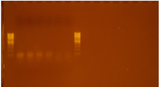
Figure 3: Showing positive bands for Leishmania tropica at 620 base pairs. Plate.1 is a Gel documentation which reveals that, Leishmania tropica bands appeared at 620 base pairs.
Gel documentation showing positive bands for Leishmania major at 670 base pairs.
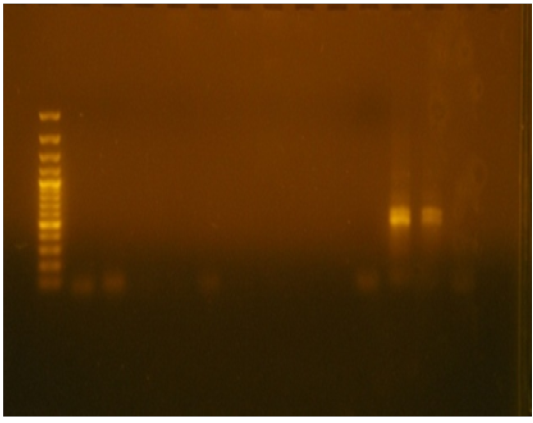
Figure 4: Showing positive bands for the Leishmania major appeared at 670 base pairs also reported similar bands of Polymerase Chain Reaction (PCR) product with sizes of 620 base pairs for Leishmania tropica and Leishmania major in Iran. Establishment of Leishmaniasis is associated with that, of the establishment of sandfly population in Sindh-Province of Pakistan. Sandfly is distributed in rural areas of Sindh-Province which are located near Indus River.
Table 1: District wise infection percentage in humans detected through Polymerase Chain Reaction (PCR).
| District | Humans | Prevalence | |
| Observed | Infected | ||
| Hyderabad | 18 | 15 | 83.33 |
| Dadu | 41 | 39 | 95.12 |
| Jamshoro | 10 | 08 | 80.00 |
| Total | 70 | 61 |
87.14 |
Total 70 samples were collected from three different districts of Sindh-Province where, 61 (87.14%) cases were suspected for Cutaneous Leishmaniasis 41% cases belong to district Dadu where as 18 % cases belong to Hyderabad and 10% cases belong to district Jamshoro.
DISCUSSION
Correct diagnosis of Leishmania species is essential to determine the clinical prognosis and a species specific therapeutic approach. The Polymerase Chain Reaction (PCR) technique has opened new windows in the diagnosis of Cutaneous Leishmaniasis, and several approaches have been developed during the last two decades, (Ronger et al., 2009; Shaheen et al., 2007). Accurate identification of the Leishmania species seems to be necessary for a variety of clinical and epidemiological reasons for deciding distinct treatment regimens and also designing appreciate control program, (Guerra et al., 2001). DNA-based techniques have commonly been used as potential tools. During this study, Cutaneous Leishmaniasis was detected in 61 (87.14 %) tissues samples out of 70 samples in three different districts of Sindh province. Our findings are not in agreement with those of which may be due to different study area (Ronger et al., 2009).
During present study we found that, Leishmania tropica and Leishmania major were predominant for Leishmania species (Soraya et al., 2010), this difference may be attributed due to geographical location which plays an important role strain zoogeography of species and strains (Rassi et al., 2013). During present study, it was seen that, exposure to various body parts also affects rate of infestation and number of infection (Daniela et al., 2012; Seray et al., 2013). Present study shows that, the infection rate for Cutaneous Leishmaniasis through different body parts was 51.42% on face area 22.85% on hand, 18.57 % on leg and 7.14% on abdomen reported 54%, 33% and 13% lesions of Leishmaniasis on face, hands and legs respectively (Aneela et al., 2011; Arfan et al., 2011). Polymerase Chain Reaction (PCR) has provided the ability to diagnose and identify Leishmania species. Polymerase Chain Reaction (PCR) based-assays have been found significantly sensitive then the histopathological methods also used for further diagnosis (Grazielley et al., 2012; Mormano et al., 2013), Establishment of Leishmaniasis is associated with that, of the establishment of sand fly population. In Sindh, Province of Pakistan, sand fly is distributed in those districts which are located on right bank of river Indus (Fallah et al., 2011, Ershadi et al., 2012, Seray et al., 2013).
Present study covered cases from three districts viz. Dadu, Jamshoro and Hyderabad. As matter of fact that, sand flies population is much distributed in districts, Dadu and Jamshoro as they share their borders with, Baluchistan-Province where population of sand flies is well established. In fact, data on Leishmaniasis in district, Hyderabad does not actually mean that, Hyderabad has high intensity of infection. More cases were seen in district, Hyderabad for the reason that, for interior Sindh, Hyderabad is hub of, Medical facilities, where people from other districts come for medical treatment.
CONCLUSION
Leishmaniasis was discovered 100 years ago but disease has not been eradicated largely organized and collaborated efforts are requires attention of health authorities to take appropriate measures for its effective control, failing which; may create more than 80% prevalence to public health problems. Cutaneous Leishmaniasis is endemic and spreading mostly found in rural areas of Sindh-Province and their surrounded areas. The disease caused by the intracellular protozoan parasite Leishmania represent a major global health problem and World Health Organization (WHO) classified neglected tropical disease. Nearly 10% of the world’s population is at risk of acquiring a form of Leishmaniasis. Worldwide it is estimated that there are 12 million active cases of Leishmaniasis, with 2 million new cases occurring each year. Among parasitic infections, this disease is responsible for the highest number of disability adjusted life years a measure of health burden after malaria.
Acknowledgments
The authors highly acknowledged the Department of Veterinary Microbiology, Faculty of Animal Husbandry & Veterinary Sciences Sindh Agriculture University Tandojam, Department of Veterinary Parasitology, Faculty of Animal Husbandry & Veterinary Sciences, Sindh Agriculture University Tandojam and Central Veterinary Diagnostic Laboratory (CVDL) Tandojam, Sindh-Pakistan for providing research facilities to carry out some part of this work and their inputs and guidelines during this manuscript.
CONFLICT OF INTEREST
The authors declare no conflict of interest.
AUTHORS CONTRIBUTION
Muhammad Ismail Qureshi was conducted the research work whereas, Abdul Ahad Soomro was the advisor. Muhammad Ismail Qureshi wrote the manuscript.
REFERENCES
