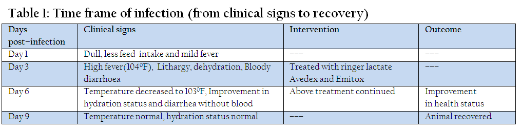
Table 1: Time frame of infection (from clinical signs to recovery)
Shumaila Manzoor*, Aatka Jamil
*Corresponding author:shamaleo@hotmail.com
ARTICLE CITATION:
Manzoor S and Jamil A (2013). Canine parvovirus associated with bloody diarrhea in Labrador retriever pup. Res. J. Vet. Pract. 1 (3): 34 – 35.
Received: 2013–08–30, Revised: 2013–09–16, Accepted: 2013–09–18
The electronic version of this article is the complete one and can be found online at
(
http://nexusacademicpublishers.com/table_contents_detail/13/74/html
)
which permits unrestricted use, distribution, and reproduction in any medium, provided the original work is properly cited
This report is about the canine parvovirus which affects mostly young dogs. A Labrador retriever pup of 3 months was presented with the complaint of bloody diarrhea and decreased feed intake. On physical examination, animal was lethargic, depressed and dehydrated. There was no history of de–worming and vaccination. As the parvovirus mostly infects young pups and is associated with diarrhea, dehydration and anorexia so the current infection was diagnosed as parvovirus. As there is no precise treatment of viral diseases in our situation so the pup was treated symptomatically using Ringer Lactate infusion @ 15ml/kg of body weight along with injection emitox @ 2ml and injection Avedex @ 2ml, initially for three days. Prognosis of disease is usually poor but fortunately pup was improving and was quite better on day third onward. So the same treatment was continued for next three days except Ringer Lactate infusion and animal recovered 7 days post–infection.
Canine parvovirus type 2 (CPV–2) is a small, non–enveloped DNA virus that emerged suddenly in 1978 as an enteric pathogen of dogs. Two antigenic variants, CPV–2a and CPV–2b, are now distributed worldwide (Truyen, 2006).
Canine parvovirus infection is an acute viral disease of dogs characterized by vomiting and diarrhea especially in pups (Pollock & Carmichael, 1983). Zafar et al., (1999) also studied that physical examination findings can include anorexia, depression, vomiting and watery bloody diarrhea. Clinical signs usually develop within 3–7 days of infection in case of parvoviral enteritis.
As a result, diseased animals face a much loss in their body weight. The disease is usually prevalent in non–vaccinated dogs due to unawareness of the owners, high costs of vaccines, poor husbandry and biosecurity practices. Constant presence of pathogens at particular places makes the disease endemic for that picky area (William et al., 2002).
The disease is also characterized by hemorrhagic enteritis, bloody diarrhea and myocarditis in young pups (Panda et al., 2009).A Labrador retriever pup of three months, was presented at Civil Veterinary Hospital, Hazro, district Attock, Punjab, with the complaint of bloody diarrhea for last 3 days. There was decreased feed and water intake. Another pup of the owner died within seven days after the onset of similar symptoms. There was no history of de–worming and vaccination.
On physical examination, there was fever (1040F) and animal was dehydrated. Animal was straining and was lethargic & depressed. Based on physical examination and keeping the age of animal in mind along with complaint of bloody diarrhea, it was diagnosed that animal was suffering from Canine Parvovirus. Following treatment was given for six days and animal recovered with a clinical phase of disease for ten days (Table 1).
Owner was advised to keep the animal hydrated by offering ice cubes or other oral rehydrating solutions. Also advised to keep the unhealthy pup isolated from the healthy one and clean the area where these pups were kept. Vaccinate the healthy pup as soon as possible.
Canine parvovirus (CPV) is a highly contagious and a common cause of acute, infectious gastrointestinal illness in pups. The disease is more common in pups ranging from 1–5 months of age. Lack of maternal immunity and poor immuno–competency is to be the cause of high incidence (Muzaffar et al., 2006).
Since the late 1970s, the CPV–2 strain has been known to be a major etiologic agent of infectious gastroenteritis in young dogs (Appel et al., 1979; Morais and Costa, 2007). Thus, canine parvovirus, which occurs with high frequency and endures for long periods of time in the environment, has stood out among other diseases because it results in high rates of morbidity and mortality (Hoskins, 1997). In untreated cases death occurs within 3 days (Muzaffar et al., 2006).
Prevalence percentage of canine parvovirus is very less reported than the reality due to less case reporting. Saghir Ahmed and Masood Rabbani (1999) studied that prevalence of canine parvovirus was 21.33 percent in Pakistan.
Canine parvo is commonly observed in dog breeds like German shepherd (84%), in Rottweiller (9%), in Doberman (5%) and in Bull terriers (2%). The disease is not recorded in dogs with non–descriptive breeds (Muzaffar et al., 2006). Factors contributing to the manifestations of CPV infection are multiple with elements of host, pathogen, secondary infections, fundamental stressors and environment affecting brutality and outcome (Schoeman et al., 2013)
The main reason for the high incidence of the disease is carelessness in the preventive measures. It is the need of time to build awareness among people about the preventive vaccination of dogs as some of their diseases could be zoonotic.
Authors want to acknowledge Dr. Muhammad Abubakar (Scientific Officer, National Veterinary Laboratories, Islamabad) for his inspiring and encouraging support. Thanks are also payable to the Veterinary staff at Hazro Clinic for their cooperation and providing the help.
Appel MJG, Cooper BJ, Greisen H, Scott FW and Carmichael LE (1979). Canine viral enteritis. I. Status report on corona and parvo–like viral enteritides. Cornell Vet.69: 123–133.
PMid:223812
Hoskins JD (1997). Update on canine parvoviral enteritis. Vet. Med. 92: 694–709.
Jafri SA and Rabbani M (1999). Prevelance of canine diseases in Lahore area. Pakistan Vet. J. 19: 40–42.
Morais MP and Costa PR (2007). Parvoviridae. In: Flores, E.F. (Ed.), Virologia Veterinária. Ed. da UFSM, Santa Maria, pp. 388–392.
Muzaffar AK, Rabbani M, Muhammad K, Murtaza N and Nazir J (2006). Isolation and characterization of canine parvovirus. Int. J. Agri. Biol. 8: 898–900.
Panda D, Patra RC, Nandi S and Swarup D (2009). Oxidative stress indices in gastroenteritis in dogs with canine parvoviral infection. Res. Vet Sci. 86(1):36–42.
http://dx.doi.org/10.1016/j.rvsc.2008.05.008
PMid:18572211
Pollock RV and Carmichael LE (1983). Canine viral enteritis. Vet. Clin. North. American Small Anim. Pract. 13: 551–66.
PMid:6316616
Schoeman JP, Goddard A and Leisewitz AL (2013). Biomarkers in canine parvovirus enteritis. N. Z. Vet J. 61(4):217–22.
http://dx.doi.org/10.1080/00480169.2013.776451
PMid:23477413
Truyen U (2006). Evolution of canine parvovirus—a need for new vaccines? Vet Microbiol.;117:9–13.
http://dx.doi.org/10.1016/j.vetmic.2006.04.003
PMid:16765539
William A, Chaudhari SUR and Atsanda NN (2002). Prevalence of some diseases of dogs and cats at the state government veterinary clinic in Maiduguri (Nigeria). Pakistan Vet. J. 22: 56–8.
Zafar MS, Khan SA and Rabbani A (1999). Hematological studies and estimation of electrolytes in dogs. Pakistan Vet. J. 19(1): 40–42.